
French Kidney Cancer Organoid Study Links Culture Models with the UroCCR Cohort
A Caen feasibility study compared seven culture conditions for clear cell renal cell carcinoma. The model collection was connected with a clinical registry, while tissue characterization revealed variable marker preservation.
Building a Model Collection Alongside Clinical Data
Researchers at Caen University Hospital and the University of Caen Normandy evaluated whether renal tumor organoids could be generated within the UroCCR clinical research framework. The study linked surgical samples, matched histology and organoid cultures with a structured clinical database, creating a basis for future laboratory–clinical comparisons in clear cell renal cell carcinoma.
Eighteen patients entered the sampling study. The investigators reported 12 established tumor cultures, and subsequent clear cell carcinoma analyses focused on the 12 cases with confirmed histology. Five of the twelve cultures underwent histological characterization, corresponding to 41.7%.
Comparing Culture Conditions and Durability
Seven conditions varied the basal medium, extracellular matrix and growth-factor supplementation. The comparison included Matrigel or related basement membrane matrix and a collagen-based air–liquid interface approach. Some cultures were started directly from surgical material, while others came from cryopreserved samples.
Five conditions supported growth. The condition containing EGF, FGF, heparin and B27 was the best-performing condition in 5 of 12 cases, but no single medium worked for every sample. Across the collection, the reported median culture duration was 52 days and the mean number of passages was two. Some cultures persisted longer, while others stopped much earlier.
The air–liquid interface cultures did not sustain growth beyond the first week. The investigators described difficulty observing structures through opaque collagen, disruption during passaging and limited starting material shared across several parallel conditions. These observations show how a format that is useful in other tumors can encounter practical problems in a particular collection.
Comparing the Cultures with Their Source Tumors
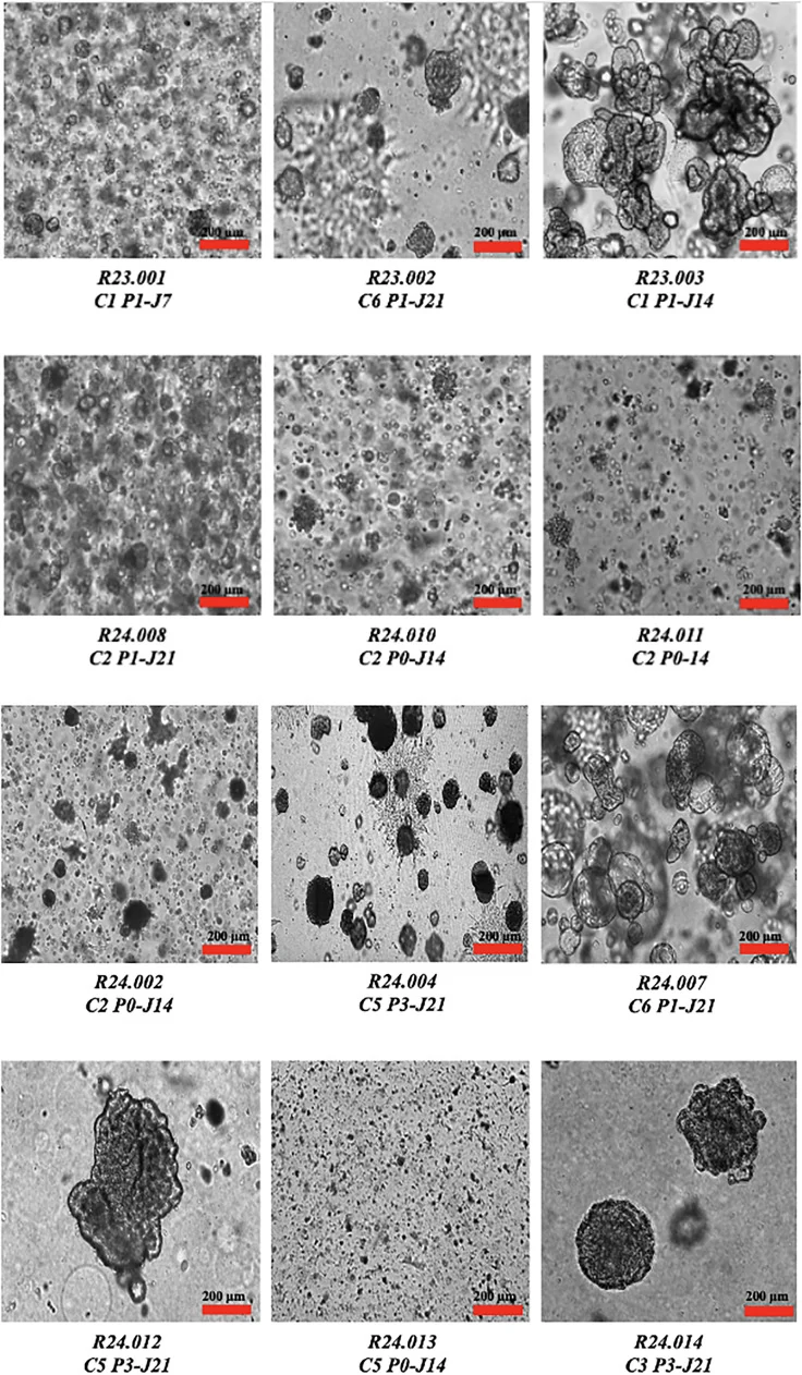
Microscopy showed cystic and solid structures. Some cystic organoids increased in size between passages, while solid structures declined in number. Growth was monitored qualitatively rather than through a formal standardized growth-curve analysis.
Five cultures were assessed histologically. They retained several features of the corresponding tumors, but staining for CA-IX and CK7 varied. CK7 was positive in some organoids whose source tumors were negative, and one culture had insufficient material for the full marker comparison. Those differences matter when deciding how faithfully a model represents the source tumor after time in culture.
A Feasibility Resource Before Drug-Response Validation
The principal contribution was the connection between patient-derived cultures and the UroCCR clinical infrastructure, with parallel tissue samples for comparison. The reported study did not perform genomic or transcriptomic validation, and it did not establish a drug-response relationship with patient outcomes. Therapeutic screening remained a proposed use of the collection.
The cultures also lacked a characterized immune and vascular compartment, which is relevant to renal cancer therapies acting through those systems. Improving growth consistency, reducing the time to characterization and preserving material for matched analysis are the immediate questions raised by the study. The registry link provides a framework in which those improvements could later be tested against clinical data.